Symptoms Of Bursitis Of The Feet
Overview
Calcaneal bursitis is seen somewhat frequently, particularly if the clinician has a predominantly musculoskeletal practice. No mortality is associated with calcaneal bursitis. Morbidity is associated with progressive pain and limping (antalgic gait) in patients who have not received adequate treatment. If chronic inflammation also affects the distal Achilles tendon, rupture of the tendon may occur. No race predilection has been documented. Calcaneal bursitis is observed in men and women. However, some increased risk may be incurred by women who wear high-heeled shoes. Calcaneal bursitis is commonly observed in middle-aged and elderly persons, the condition is also seen in athletes of all ages.Bursitis is a painful swelling of a small sac of fluid called a bursa. Bursae (plural of bursa) cushion and lubricate areas where tendons, ligaments, skin, muscles, or bones rub against each other. People who repeat the same movement over and over or who put continued pressure on a joint in their jobs, sports, or daily activities have a greater chance of getting bursitis.
Causes
Bursitis is commonly caused by overuse and repeated movements. These can include daily activities such as using tools, gardening, cooking, cleaning, and typing at a keyboard. Long periods of pressure on an area. For example, carpet layers, roofers, or gardeners who work on their knees all day can develop bursitis over the kneecap. Aging, which can cause the bursa to break down over time. Sudden injury, such as a blow to the elbow. Bursitis can also be caused by other problems, such as arthritis or infection (septic bursitis).
Symptoms
Nagging ache and swelling in or around a joint. Painful and restricted movement in the affected joint. Pain radiating into the neck or arms when bursitis strikes the shoulder (the most common site). Fever, when associated with an infection.
Diagnosis
Your GP or therapist will be able to diagnose you by both listening to your history and examining you. No X-rays or further investigation should be needed to confirm diagnosis but may be requested to check for any underlying health conditions that may have triggered the bursitis.
Non Surgical Treatment
You should rest from all activities that cause pain or limping. Use crutches/cane until you can walk without pain or limping. Ice. Place a plastic bag with ice on the foot for 15-20 minutes, 3-5 times a day for the first 24-72 hours. Leave the ice off at least 1 1/2 hours between applications. Compression. Lightly wrap an elastic bandage from the toes to mid calf, using even pressure. Wear this until swelling decreases. Loosen the wrap if your toes start to turn blue or feel cold. Elevate. Make sure to elevate the ankle above heart level. To improve symptoms of plantar calcaneal bursitis after the acute phasetry the baked bean tin stretch, using a baked bean tin roll the foot backwards and forwards. 2 minutes in the morning before putting the foot to the floor. 5-10 minutes every evening. Contrast foot baths. 10 minutes warm water. 10 minutes cool water morning and evening (morning may be missed if time is restricted). Stretches. Start with 10 stretches per day, holding the stretch for 30 seconds, then relax and then repeat. Continue this stretch daily until you can no longer feel it pulling on the heel, then progress to stretch. Do 10 per day holding for 30 seconds per stretch. When you can no longer feel it pulling on the heel proceed to stretches. Do 10 per day holding for 30 seconds on every stretch.
Surgical Treatment
Surgery to remove the damaged bursa may be performed in extreme cases. If the bursitis is caused by an infection, then additional treatment is needed. Septic bursitis is caused by the presence of a pus-forming organism, usually staphylococcus aureus. This is confirmed by examining a sample of the fluid in the bursa and requires treatment with antibiotics taken by mouth, injected into a muscle or into a vein (intravenously). The bursa will also need to be drained by needle two or three times over the first week of treatment. When a patient has such a serious infection, there may be underlying causes. There could be undiscovered diabetes, or an inefficient immune system caused by human immunodeficiency virus infection (HIV).
Prevention
Do not run if you have pain. When you begin running again, avoid running fast uphill or downhill until the tendon is fully healed. Start exercising when caregivers say that it is OK. Slowly start exercise such as bicycling when caregivers say it is OK. When doing exercises that put pressure on the ankles, such as running or walking, exercise on flat, even surfaces. Avoid doing these exercises on very hard surfaces such as asphalt or concrete. Stretch before exercising. Always warm up your muscles and stretch gently before exercising. Do cool down exercises when you are finished. This will loosen your muscles and decrease stress on your heel. Wear heel protectors. Use soft foam or felt heel pads (wedges or cups) to help decrease pressure against your heel. Ask your caregiver which heel pads are the best for you. Wear well-fitting shoes. Buy running or exercise shoes that support and fit your feet well. Do not wear low-cut shoes. Talk to your caregiver or go to a special exercise footwear store to get well-fitting athletic shoes. Ask your caregiver if you should wear specially-made shoe inserts called orthotics (or-THOT-iks). Orthotics can line up your feet in your shoes to help you run, walk and exercise correctly.
How To Help Hammertoe Pain
 Overview
The smallest four toes of each foot have three bony segments connected by two joints. Hammertoe is a deformity in which one or more of the small toes develops a bend at the joint between the first and second segments. The tip of the toe turns downward, making it look like a hammer or claw. The second toe is the one most often affected. hammertoe may hammertoe be more likely to occur when the second toe is longer than the first toe or when the arch of the foot is flat.
Causes
A common cause of hammertoe and mallet toe is wearing improper footwear - shoes that are too tight in the toe box, or high-heel shoes. Wearing shoes of either type can push your toes forward, crowding one or more of them into a space that's not large enough to allow your toes to lie flat. Hammertoe and mallet toe deformities can also be inherited and may occur despite wearing appropriate footwear. The result is a toe that bends upward in the middle and then curls down in a hammer-like or claw-like shape. Your shoes can rub against the raised portion of the toe or toes, causing painful corns or calluses. The bottom of the affected toe can press down, creating the mallet-like appearance of mallet toe. At first, a hammertoe or mallet toe may maintain its flexibility and lie flat when you're not wearing crowded footwear. But eventually, the tendons of the toe may contract and tighten, causing your toe to become permanently stiff.
 Symptoms
Patients with hammer toe(s) may develop pain on the top of the toe(s), tip of the toe, and/or on the ball of the foot. Excessive pressure from shoes may result in the formation of a hardened portion of skin (corn or callus) on the knuckle and/or ball of the foot. Some people may not recognize that they have a hammer toe, rather they identity the excess skin build-up of a corn.The toe(s) may become irritated, red, warm, and/or swollen. The pain may be dull and mild or severe and sharp. Pain is often made worse by shoes, especially shoes that crowd the toes. While some hammer toes may result in significant pain, others may not be painful at all. Painful toes can prevent you from wearing stylish shoes.
Diagnosis
Most health care professionals can diagnose hammertoe simply by examining your toes and feet. X-rays of the feet are not needed to diagnose hammertoe, but they may be useful to look for signs of some types of arthritis (such as rheumatoid arthritis) or other disorders that can cause hammertoe. If the deformed toe is very painful, your doctor may recommend that you have a fluid sample withdrawn from the joint with a needle so the fluid can be checked for signs of infection or gout (arthritis from crystal deposits).
Non Surgical Treatment
People with a hammer toe benefit from wearing shoes in which the toe box is made of a flexible material and is wide enough and high enough to provide adequate room for the toes. High-heeled shoes should be avoided, because they tend to force the toes into a narrow, flat toe box. A doctor may recommend an insert (orthotic) for the shoe to help reduce friction and pressure on the hammer toe. Wearing properly fitted shoes may reduce pain and inflammation. It may also prevent ulcers from developing and help existing ulcers heal. However, the hammer toe does not disappear.
Surgical Treatment
Surgery to correct for a hammertoe may be performed as a day procedure. There are several different types of procedures that can be used depending on the foot structure and if the deformity is flexible or rigid.
Bunions Causes And Cures
Overview
 Bunion pain can be debilitating and bunion surgery can be a scary decision most people would rather put off. I know. I've been there and I've learned that IF you know what to expect ahead of time and what questions to ask your doctor, you will lessen your fear, make better decisions, and increase your success with bunion surgery and bunion surgery recovery. No matter how good your podiatrist or bunion surgeon is, the truth is, no one knows exactly what's right for you except you and that's what can make your decision so scary. What if the surgery doesn't really fix the problem, and like others you read about or hear about, you end up worse off than before? It's not always clear what to do, but with the right information, questions, and support, it can be an easier decision for you.
Causes
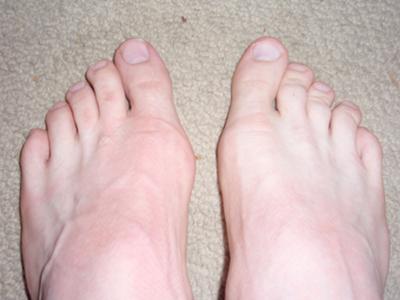
Prolonged pressure against the inside portion of the 1st MTP joint can lead to Bunions. This most commonly occurs as a result of wearing pointed toe shoes or shoes that are too tight. Another cause is over-pronation. Normal ?toe-off?occurs from bottom of the big toe. Over-pronation can cause one to ?toe-off? on the inside portion of the big toe versus the bottom. Over time, there is a retrograde force into the joint which pushes it out stretches the joint capsule. This tearing and stretching of the joint capsule as well as the wear and tear on the cartilage is what causes the pain.
Symptoms
The symptoms of bunions include irritated skin around the bunion, pain when walking, joint redness and pain, and possible shift of the big toe toward the other toes. Blisters may form more easily around the site of the bunion as well. Having bunions can also make it more difficult to find shoes that fit properly; bunions may force a person to have to buy a larger size shoe to accommodate the width the bunion creates. When bunion deformity becomes severe enough, the foot can hurt in different places even without the constriction of shoes because it then becomes a mechanical function problem of the forefoot.
Diagnosis
When an x-ray of a bunion is taken, there is usually angulation between the first metatarsal bone and the bones of the big toe. There may also be angulation between the first and second metatarsal bones. These angular irregularities are the essence of most bunions. In general, surgery for bunions aims to correct such angular deformities.
Non Surgical Treatment
Some bunions can be treated without surgery. If you have a bunion, wear shoes that are roomy enough so that they won?t put pressure on it. You can choose to have your shoes stretched out professionally or try cushioning the painful area with protective pads. Orthotics have been shown to help prevent progression of bunions. Oral nonsteroidal anti-inflammatory drugs (NSAIDs), such as ibuprofen, may be recommended to reduce pain and inflammation. Applying an ice pack several times a day can also help reduce inflammation and pain. If your bunion progresses to a point where you have difficulty walking or experience pain even with accommodative shoes, surgery may be necessary. 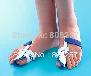
Surgical Treatment
If non-surgical treatments have failed to relieve your bunion pain, or when the pain is interfering with your daily activities, contact the Dallas bunion surgery specialists at North Texas Foot & Ankle to discuss surgical options. There are a several ways to perform bunion surgery. The best procedure for one person is not necessarily the best for another. Some procedures allow you to walk much sooner, reducing the need for crutches. Depending on your foot type, the procedure can have a greater risk for return of the bunion deformity. Other procedures may require you to be on crutches for a few weeks - but could offer a better result in the long-term. Learn more about the different types of bunion surgery.
Prevention
To help prevent bunions be sure your shoes don't cramp or irritate your toes. Choose shoes with a wide toe box - there should be space between the tip of your longest toe and the end of the shoe. Your shoes should conform to the shape of your feet without squeezing or pressing any part of your foot. Avoid pointy-toed shoes.
Overpronation Cause And Treatment
Overview
Overpronation is a condition in which the foot rolls excessively down and inward. The arch may elongate and collapse (or ?fall?) and the heel will lean inward. Overpronation should not be confused with pronation. Pronation is a normal motion of the foot during weight bearing and allows the foot to absorb shock as it contacts the ground.
Causes
Congenital "Flat Feet" - an individual may be born with feet that lack an appropriately supportive arch thereby predisposing the individual to this foot condition. Excessive Weight (Obesity) Too much weight on the foot from either obesity or pregnancy may be a factor. Repetitive Impact walking on flat, hard surfaces continuously places unnatural stress on the foot arch.
Symptoms
Common conditions that develop with prolonged overpronation typically include plantar fasciitis, achilles tendonitis, shin splints, posterior tibial stress syndrome and even IT band syndrome. With long term neglect you may see the development of bunyons, foot deformities and early onset of hip and knee arthritis.
Diagnosis
Pronounced wear on the instep side of shoe heels can indicate overpronation, however it's best to get an accurate assessment. Footbalance retailers offer a free foot analysis to check for overpronation and help you learn more about your feet.
Non Surgical Treatment
Over-Pronation can be treated conservatively (non-surgical treatments) with over-the-counter orthotics. These orthotics should be designed with appropriate arch support and medial rearfoot posting to prevent the over-pronation. Footwear should also be examined to ensure there is a proper fit. Footwear with a firm heel counter is often recommended for extra support and stability. Improperly fitting footwear can lead to additional foot problems.
Surgical Treatment
Subtalar Arthroereisis. Primary benefit is that yje surgery is minimally invasive and fully reversible. the primary risk is a high chance of device displacement, generally not tolerated in adults.
An implant is pushed into the foot to block the excessive motion of the ankle bone. Generally only used in pediatric patients and in combination with other procedures, such as tendon lengthening. Reported removal rates vary from 38% - 100%, depending on manufacturer.
Physiotherapy And Severs Disease
Overview
The muscle group at the back of the lower leg is commonly called the calf. The calf comprises of 2 major muscles (known as the gastrocnemius and soleus) both of which insert into the heel bone via the Achilles tendon. In people who have not yet reached skeletal maturity, a growth plate exists where the Achilles tendon inserts into the heel bone. This growth plate is primarily comprised of cartilage. Every time the calf contracts, it pulls on the Achilles tendon which in turn pulls on the heel's growth plate. When this tension is too forceful or repetitive, irritation to the growth plate may occur resulting in pain and sometimes an increased bony prominence at the back of the heel. This condition is called Severs disease. Severs disease is typically seen in children or adolescents during periods of rapid growth. This is because muscles and tendons become tighter as bones become longer. As a result, more tension is placed on the heel's growth plate.
Causes
Sever?s disease is often an overuse injury. It can be caused by playing sports, especially those involving high impact. Sever?s disease can also be linked to growth which can place a stress on the attachment of the Achilles tendon.
Symptoms
Sever's Disease is most commonly seen in physically active girls and boys from ages 10 to 15 years old. These are the years when the growth plate is still ""open,"" and has not fused into mature bone. Also, these are the years when the growth plate is most vulnerable to overuse injuries, which are usually caused by sports activities. The most common symptoms of this disease include. Heel pain in one or both heels. Usually seen in physically active children, especially at the beginning of a new sports season. The pain is usually experienced at the back of the heel, and includes the following areas. The back of the heel (that area which rubs against the back of the shoe). The sides of the heel. Actually, this is one of the diagnostic tests for Sever's Disease, squeezing the rear portion of the heel from both sides at the same time will produce pain. It is known as the Squeeze Test.
Diagnosis
In Sever's disease, heel pain can be in one or both heels. It usually starts after a child begins a new sports season or a new sport. Your child may walk with a limp. The pain may increase when he or she runs or jumps. He or she may have a tendency to tiptoe. Your child's heel may hurt if you squeeze both sides toward the very back. This is called the squeeze test. Your doctor may also find that your child's heel tendons have become tight.
Non Surgical Treatment
The immediate goal of treatment is pain relief. Because symptoms generally worsen with activity, the main treatment for Sever's disease is rest, which helps to relieve pressure on the heel bone, decreasing swelling and reducing pain. As directed by the doctor, a child should cut down on or avoid all activities that cause pain until all symptoms are gone, especially running barefoot or on hard surfaces because hard impact on the feet can worsen pain and inflammation. The child might be able to do things that do not put pressure on the heel, such as swimming and biking, but check with a doctor first.
What Are The Major Causes Of Posterior Tibial Tendon Dysfunction ?
Overview
There are some things that gain value as they age. Antique dealers are always on the lookout for pieces that have a certain ?wear and tear? look that will bring a high price tag. Our feet on the other hand, don?t always fair as well when they have experienced a lot of wear and tear. Cumulative stress and impact can cause your foot structure to weaken and become prone to injury, especially when you have a flat foot. This is the case with a condition called posterior tibial tendon dysfunction. 
Causes
Posterior tibial tendon dysfunction is the most common cause of acquired adult flatfoot. Sometimes this can be a result of specific trauma, but usually the tendon becomes injured from wear and tear over time. This is more prevalent in individuals with an inherited flat foot but excessive weight, age, and level of activity are also contributing factors.
Symptoms
Pain along the inside of the foot and ankle, where the tendon lies. This may or may not be associated with swelling in the area. Pain that is worse with activity. High-intensity or high-impact activities, such as running, can be very difficult. Some patients can have trouble walking or standing for a long time. Pain on the outside of the ankle. When the foot collapses, the heel bone may shift to a new position outwards. This can put pressure on the outside ankle bone. The same type of pain is found in arthritis in the back of the foot. Asymmetrical collapsing of the medial arch on the affected side.
Diagnosis
First, both feet should be examined with the patient standing and the entire lower extremity visible. The foot should be inspected from above as well as from behind the patient, as valgus angulation of the hindfoot is best appreciated when the foot is viewed from behind. Johnson described the so-called more-toes sign: with more advanced deformity and abduction of the forefoot, more of the lateral toes become visible when the foot is viewed from behind. The single-limb heel-rise test is an excellent determinant of the function of the posterior tibial tendon. The patient is asked to attempt to rise onto the ball of one foot while the other foot is suspended off the floor. Under normal circumstances, the posterior tibial muscle, which inverts and stabilizes the hindfoot, is activated as the patient begins to rise onto the forefoot. The gastrocnemius-soleus muscle group then elevates the calcaneus, and the heel-rise is accomplished. With dysfunction of the posterior tibial tendon, however, inversion of the heel is weak, and either the heel remains in valgus or the patient is unable to rise onto the forefoot. If the patient can do a single-limb heel-rise, the limb may be stressed further by asking the patient to perform this maneuver repetitively.
Non surgical Treatment
Depending on the stage of the deformity and patient?s functional goals, various treatment options are available. Some patients improve with conservative care which includes rest and immobilization, shoe modifications, orthoses and bracing, or physical therapy. Surgery might be warranted for advanced stages of the condition. Often a combination of procedures including tendon and muscle augmentation, tendon transfers, realigning of bones or fusion of certain joints might be necessary in more advanced cases. Your doctor will evaluate and recommend an individualized plan of care with your specific needs in mind. 
Surgical Treatment
In cases of PTTD that have progressed substantially or have failed to improve with non-surgical treatment, surgery may be required. For some advanced cases, surgery may be the only option. Your foot and ankle surgeon will determine the best approach for you.
Heel Soreness The Causes, Warning Signs And Therapy Methods
Overview

The most common form of Heel Pain, is pain on the bottom of the heel. It tends to occur for no apparent reason and is often worse when first placing weight on the foot. Patients often complain of pain the first thing in the morning or after getting up to stand after sitting. The pain can be a sharp, searing pain or present as a tearing feeling in the bottom of the heel. As the condition progresses there may be a throbbing pain after getting off your feet or there may be soreness that radiates up the back of the leg. Pain may also radiate into the arch of the foot.
Causes
Heel pain can have many causes but the vast majority is caused by plantar fasciitis. Plantar means, ?bottom of the foot.? Fascia is a ligament or ?bundle? of ligaments. The plantar fascia is the thick ligament that helps to hold up the foot and provide spring in our step. Plantar fasciitis is an inflammation of the plantar fascia and causes more than 90% of heel pain among adults in the US. Plantar fasciitis can be acute, that is, as simple strain of the ligament but often is chronic, hanging on for months if not years. Why does that happen? The answer is poor foot mechanics, the foot sinking down too far allowing the plantar fascia to overstretch with each step taken.
Symptoms
The primary symptom is pain in the heel area that varies in severity and location. The pain is commonly intense when getting out of bed or a chair. The pain often lessens when walking.
Diagnosis
To arrive at a diagnosis, the foot and ankle surgeon will obtain your medical history and examine your foot. Throughout this process the surgeon rules out all the possible causes for your heel pain other than plantar fasciitis. In addition, diagnostic imaging studies such as x-rays or other imaging modalities may be used to distinguish the different types of heel pain. Sometimes heel spurs are found in patients with plantar fasciitis, but these are rarely a source of pain. When they are present, the condition may be diagnosed as plantar fasciitis/heel spur syndrome.
Non Surgical Treatment
If the plantar fasciitis is acute, that is, a sprain of the plantar fascia then it is basically treated as a sprain, with anti-inflammatory drugs, ice, rest, possibly physical therapy. If chronic, the poor foot mechanics need be addressed. Foot mechanics are changed by use of specially moulded shoe inserts known as orthotics. Someone with plantar fasciitis needs an orthotic designed to relieve strain on the plantar fascia. Orthotics are often confused with arch supports. Arch supports, by holding up the arch can remove some of the tension from the plantar fascia. Orthotics, on the other hand, do most of their work on the heel and ball of the foot repositioning the foot for maximized function. What can you do before you see the foot doctor? First, try doing your own version of deep tissue massage by rolling a frozen cola bottle or can from the heel forward into the arch. Do it gently. Do stretching but the key to good stretching is not to stretch too hard so generally avoid weight bearing (standing) stretches but sit on a soft surface like your bed and pull the foot backward on the leg as far as it will go, holding for 20 seconds and relaxing for 5 seconds. Each 25 second ?set? can be repeated 5 times and you have invested about 2 minutes in giving yourself a lot of help. Watch out for the shoes you wear. It is tempting to obtain shoes that are colorful and soft. Here is the proof that soft shoes are bad. Wrap a pillow around your foot with duct tape and walk for a block or two. You will come back with your foot hurting more because your foot sank down deeper into the soft surface, allowing the ligament to stretch more. The shoes should be stiff in the shank and flexible at the ball. Such shoes, to running buffs, are known as motion control shoes or stability shoes so going to one of the small specialty running shoes stores is a good place to start. If you don?t have a desk job, or have an industrial job see if light duty is available. A note from your doc may be all that is required in most cases and most doctors are happy to oblige.
Surgical Treatment
When a diagnosis of plantar fasciitis is made early, most patients respond to conservative treatment and don?t require surgical intervention. Often, when there is a secondary diagnosis contributing to your pain, such as an entrapped nerve, and you are non-responsive to conservative care, surgery may be considered. Dr. Talarico will discuss all options and which approach would be the most beneficial for your condition.
Prevention

Wearing real good, supportive shoes are a great way to avoid heel pain. Usually, New Balance is a good shoe to wear, just for everyday shoe gear. By wearing proper footwear and performing thorough stretches, athletes can help prevent frequent heel pain. If you are starting to get a little discomfort or pain in the feet or heel, know that pain is not normal. So if you are having pain, you should be proactive and visit our office. If you let heel pain get out of control you could run into several other problems. It is always suggested to visit a podiatrist whenever you are experiencing pain.
The most common form of Heel Pain, is pain on the bottom of the heel. It tends to occur for no apparent reason and is often worse when first placing weight on the foot. Patients often complain of pain the first thing in the morning or after getting up to stand after sitting. The pain can be a sharp, searing pain or present as a tearing feeling in the bottom of the heel. As the condition progresses there may be a throbbing pain after getting off your feet or there may be soreness that radiates up the back of the leg. Pain may also radiate into the arch of the foot.
Causes
Heel pain can have many causes but the vast majority is caused by plantar fasciitis. Plantar means, ?bottom of the foot.? Fascia is a ligament or ?bundle? of ligaments. The plantar fascia is the thick ligament that helps to hold up the foot and provide spring in our step. Plantar fasciitis is an inflammation of the plantar fascia and causes more than 90% of heel pain among adults in the US. Plantar fasciitis can be acute, that is, as simple strain of the ligament but often is chronic, hanging on for months if not years. Why does that happen? The answer is poor foot mechanics, the foot sinking down too far allowing the plantar fascia to overstretch with each step taken.
Symptoms
The primary symptom is pain in the heel area that varies in severity and location. The pain is commonly intense when getting out of bed or a chair. The pain often lessens when walking.
Diagnosis
To arrive at a diagnosis, the foot and ankle surgeon will obtain your medical history and examine your foot. Throughout this process the surgeon rules out all the possible causes for your heel pain other than plantar fasciitis. In addition, diagnostic imaging studies such as x-rays or other imaging modalities may be used to distinguish the different types of heel pain. Sometimes heel spurs are found in patients with plantar fasciitis, but these are rarely a source of pain. When they are present, the condition may be diagnosed as plantar fasciitis/heel spur syndrome.
Non Surgical Treatment
If the plantar fasciitis is acute, that is, a sprain of the plantar fascia then it is basically treated as a sprain, with anti-inflammatory drugs, ice, rest, possibly physical therapy. If chronic, the poor foot mechanics need be addressed. Foot mechanics are changed by use of specially moulded shoe inserts known as orthotics. Someone with plantar fasciitis needs an orthotic designed to relieve strain on the plantar fascia. Orthotics are often confused with arch supports. Arch supports, by holding up the arch can remove some of the tension from the plantar fascia. Orthotics, on the other hand, do most of their work on the heel and ball of the foot repositioning the foot for maximized function. What can you do before you see the foot doctor? First, try doing your own version of deep tissue massage by rolling a frozen cola bottle or can from the heel forward into the arch. Do it gently. Do stretching but the key to good stretching is not to stretch too hard so generally avoid weight bearing (standing) stretches but sit on a soft surface like your bed and pull the foot backward on the leg as far as it will go, holding for 20 seconds and relaxing for 5 seconds. Each 25 second ?set? can be repeated 5 times and you have invested about 2 minutes in giving yourself a lot of help. Watch out for the shoes you wear. It is tempting to obtain shoes that are colorful and soft. Here is the proof that soft shoes are bad. Wrap a pillow around your foot with duct tape and walk for a block or two. You will come back with your foot hurting more because your foot sank down deeper into the soft surface, allowing the ligament to stretch more. The shoes should be stiff in the shank and flexible at the ball. Such shoes, to running buffs, are known as motion control shoes or stability shoes so going to one of the small specialty running shoes stores is a good place to start. If you don?t have a desk job, or have an industrial job see if light duty is available. A note from your doc may be all that is required in most cases and most doctors are happy to oblige.
Surgical Treatment
When a diagnosis of plantar fasciitis is made early, most patients respond to conservative treatment and don?t require surgical intervention. Often, when there is a secondary diagnosis contributing to your pain, such as an entrapped nerve, and you are non-responsive to conservative care, surgery may be considered. Dr. Talarico will discuss all options and which approach would be the most beneficial for your condition.
Prevention
Wearing real good, supportive shoes are a great way to avoid heel pain. Usually, New Balance is a good shoe to wear, just for everyday shoe gear. By wearing proper footwear and performing thorough stretches, athletes can help prevent frequent heel pain. If you are starting to get a little discomfort or pain in the feet or heel, know that pain is not normal. So if you are having pain, you should be proactive and visit our office. If you let heel pain get out of control you could run into several other problems. It is always suggested to visit a podiatrist whenever you are experiencing pain.